Files
Download Full Text (72.5 MB)
Description
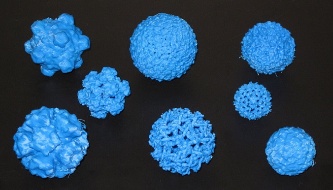
Dengue virus, database # 1THD Dengue fever, is an infectious tropical disease caused by the dengue virus and transmitted by mosquito. Symptoms include fever, headache, muscle and joint pains, and a characteristic skin rash that is similar to measles. In a small proportion of cases the disease develops into the life-threatening dengue hemorrhagic fever. Dengue is a positive strand RNA virus that is part of the flavivirus family. Dengue is an enveloped virus meaning it has a nucleocapsid protecting the RNA and the nucleocapsid is surrounded by lipids (hence the name enveloped).
Publication Date
1-18-2013
Keywords
biochemistry, icosahedron, nanobiology, pathogen, teaching, virus
Disciplines
Bacteriology | Biochemistry | Diseases | Immunology and Infectious Disease | Medical Education | Medicine and Health Sciences | Microbiology | Molecular Biology | Virology | Virus Diseases
Creative Commons License

This work is licensed under a Creative Commons Attribution 3.0 License.
Find in Thingiverse
Recommended Citation
Hall, David J., "Dengue virus" (2013). Infectious Pathogens. 1.
https://lux.lawrence.edu/chem_pathogen/1

Included in
Bacteriology Commons, Biochemistry Commons, Immunology and Infectious Disease Commons, Medical Education Commons, Molecular Biology Commons, Virology Commons, Virus Diseases Commons


Instructions
The capsids and nucleocapsids of viruses are only nanometers across and protect the viral genome encased within (http://en.wikipedia.org/wiki/Virus#Structure). The capsids are beautiful examples of icosahedral symmetry and are wonderful examples of self-assembly. Many nanotechnologists are now trying to replicate what nature does so well in order to deliver drugs more effectively among other things. In order to teach about virus structure and self-assembly, I took x-ray crystal structure data and models of various viruses from the VIPER database (.vdb files) (http://viperdb.scripps.edu/) and converted them to .stl files using the program, Chimera (http://www.cgl.ucsf.edu/chimera/). Instructions for converting the model are below as well as a short description of the virus. When slicing the virus, there are some errors; however, the virus prints well. Any suggestions and improvement to the .stl file is appreciated. You can read more about the structures at some of the web links listed at the end.
Convert vdb to stl file Go to the virus structural database at viperdb.scripps.edu/ Download the virus structure of your choice as a .vdb file Open the .vdb (VIPERdb) file in chimera cgl.ucsf.edu/chimera/ In order to print a virus capsid you shouldn't use the "multiscale" surfaces that appear when you open the VIPERdb file. Make a single surface for the whole virus by creating a simulated density map. Show the command line with Chimera menu Favorites / Command Line... and use molmap #0 10 sym i222 after opening your virus model. The molmap command will make a simulated density map and show a contour surface for that map. The resolution in the example is 10 Angstroms and the protein model #0 is replicated with icosahedral symmetry (with the VIPERdb coordinate system). Switch the volume step size to 1 in the volume dialog (or use command volume #0 step 1) to see the surface at full resolution. (thank you Tom Goddard for suggestions on using chimera) Save as stl file Open in replicator G Scale to appropriate size (many times very small and unseeable!). This can also make the .stl file VERY big. I have no idea why. Upload to cloud.nettfabb.com to fix stl. Sometimes the .stl is unfixable. I do not know why. Slice and print! During slicing, many errors do occur. However, the final model is quite printable and useable. More reading: virology.wisc.edu/virusworld/viruslist.php virology.wisc.edu/virusworld/tri_number.php viperdb.scripps.edu/icos_server.php?icspage=paradigm