
Files
Download
Download Full Text (17.5 MB)
Description
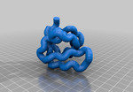
SH3 domain #1NEB. Src-homology 3 (SH3) domains bind to Pro-rich peptides that form a left-handed poly-Pro type II helix, with the minimal consensus Pro-X-X-Pro. Each Pro is usually preceeded by an aliphatic residue. Each in the aliphatic-Pro pair binds to a hydrophobic pocket on the SH3 domain.
Publication Date
1-23-2013
Keywords
biochemistry, protein, teaching
Disciplines
Biochemistry | Biochemistry, Biophysics, and Structural Biology | Bioinformatics | Life Sciences | Molecular Biology
Creative Commons License

This work is licensed under a Creative Commons Attribution 3.0 License.
Find in Thingiverse
Recommended Citation
Hall, David J., "SH3 Domain" (2013). Protein Domains. 3.
https://lux.lawrence.edu/chem_proteindomain/3
Instructions
Students learn in many different ways. One important way to learn is tactile. Some people learn best by actually touching three-dimensional models. In biochemistry classes, students usually visualize protein structures using programs such as pymol, rasmol etc. For teaching protein structure and function, I have paired 3D printed models with pymol visualization in an iPad.
There are over 40 different protein domains. The three dimensional structure of proteins and their domains is determined by x-ray crystallography or nuclear magnetic resonance (NMR). The 3D coordinates of each atom in a domain is determined and provided in a protein database (PDB) format. The PDB files for every protein structure ever determined are freely available at PDB.org. Below is an explanation of how to take a PDB file and convert it to an STL file.
Convert pdb to stl file
Select structure from pdb.org
open VMD 1.9.1 (ks.uiuc.edu/Research/vmd/)
Load pdb file →file “new molecule”
“graphics” → representations → drawing method “tube”
Radius 1.5
Resolution 10
Display →axes → off
File→ render → “enter file name” → STL → render
Open in replicator G
Scale to appropriate size (many times very small and unseeable!)
Upload to cloud.nettfabb.com to fix stl
Slice and print!